
Equivalent Text Descriptions
Universally designed documents, slides, and videos require additional preparation to make them accessible to everyone. Equivalent text descriptions (EqTD’s) provide written descriptions of graphic elements, which helps not only blind individuals, but also persons with low vision or who have cognitive or perceptual limitations.
EqTD's by Type
Diagrams |
|
|---|---|
 |
CaLT robotic training system
A simplified 2D diagram of a 3D robot-assisted gait therapy machine. The diagram includes a line drawing of a person using the machine. Various parts are labeled, and arrows indicate directional movement of some components.
|
 |
Configuration of proposed biplane fluoroscopy system
This diagrams the proposed biplane fluoroscopy setup that will allow for 3D localization. A darkened area between two x-ray sources illustrates the area where 3D localization will occur. An accompanying flow chart describes the process of integrating the bi-plane x-ray images with 3D MRI images to form the Milwaukee Foot Model.
|
 |
Example of game-playing
A screenshot of a rehabilitation video game to assist with passive range of motion shows a spaceship, a target and a random target on an outer space scene. The spaceship and random target are labeled; the spaceship represents the participant’s foot position.
|
 |
Meshed FEA model of the prototype
A computer calculated meshed FEA 3D model of a personalized orthotic prototype.
|
 |
Network Model for Home-based Units
This diagram illustrates how LAN and wireless connections are set up between a hospital server and various control units, including home based units, Bluetooth devices, the ARM9 device driver module, and the interaction with the robotic adjustable foot plate.
|
 |
Procedures for CF Population
This diagrams the numerous technologies that are integrated to complete a patient specific FEM (Finite Element Method) of a pediatric clubfoot and the use of motion analysis to optimize the conservative treatment and prevention of deformity recurrence.
|
 |
Procedures for OI Population
This diagram illustrates how various research projects (e.g. R1 and R4) within the RERC will integrate to create Patient-specific finite element methods (FEMs) of bones to assess patient-specific fracture risks. This is linked to fracture mechanisms vs. type of Osteogenesis Imperfecta as measured by two techniques in research project R1.
|
 |
Robot-Assisted Control Model
This diagram illustrates the logic and flow of information of the home based Telerehabilitation Center using a Local Autonomous Robot-assisted Control device to a hospital database. The diagram illustrates the circular flow of information from the home to the hospital database and back again to the robot-assisted home device.
|
 |
Robotic gait training system
This is a simplistic 2D diagram of a 3D robotic gait training system. The diagram includes a line drawing of a person using the machine. Various parts are labeled, and arrows indicate the directional movement of some components.
|
 |
Schematic of the Milwaukee Foot Model
This diagram shows posterior, lateral, and superior views of the foot with 6, 12, and 13 black dots, respectively, indicating the location of sensors that will be used to model the foot of individuals.
|
 |
SIMM shoulder Model
This illustration is a diagram of a dynamic model of the muscles of the shoulder.
|
 |
Top View of Foot Plate
This is a drawing of a right and left pivot-sliding foot plate for an elliptical training system. Arrows indicate the direction of movement of certain components.
|
 |
Trio-pressure measurements for flatfoot
A pressure map of the foot is illustrated using various colors to indicate different amounts of pressure.
|
 |
UE 3D inverse dynamics model
A diagram of half of a torso and one arm with variously labeled black dots indicating the location of sensors to be used to model the upper extremity dynamics of individuals.
|
Illustrations |
|
 |
3D model of pivoting and sliding mechanism
A diagram of a person using an elliptical exercise machine. Various parts color coded and labeled, and arrows indicate the direction of movement of some components, including the foot plates.
|
 |
D4 Prototype
Three pictures illustrate the construction of a foot orthosis. The left shows the hard layer, the middle the soft layer, and the right indicates the location of built-in wedges.
|
 |
VR training game exmple
This four part diagram illustrates how a person’s foot movements including sliding and pivoting, will control the actions of a character in a video game.
|
Logos |
|
 |
Children's Hospital of Wisconsin
Children's Hospital of Wisconsin logo
|
 |
Marquette University
The logo for Marquette University
|
 |
Medical College of Wisconsin
The logo for the Medical College of Wisconsin
|
 |
Milwaukee Center for Independence
Milwaukee Center for Independence logo
|
 |
Milwaukee School of Engineering
The Milwaukee School of Engineering logo
|
 |
National Institute on Disability and Rehabilitation Research
National Institute on Disability and Rehabilitation Research logo
|
 |
Rehabilitation Institute of Chicago
Logo for the Rehabilitation Institute of Chicago
|
 |
Rehabilitation Institute of Chicago
Logo for the Rehabilitation Institute of Chicago
|
 |
Shriners Hospitals for Children
Logo for Shriners Hospitals for Children
|
 |
Shriners Hospitals for Children
Logo for Shriners Hospitals for Children
|
 |
Tech4POD Mascot
This image shows a light blue teddy bear facing right and running from dot to dot on a graph.
|
 |
University of Wisconsin - Milwaukee
Logo for University of Wisconsin - Milwaukee
|
Photographs |
|
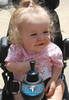 |
brianna_wheelchair
A young smiling girl in a small motorized wheelchair.
|
 |
Alexander Griffin and Poster
Alexander Griffin stands next to his poster in the Medical College Research Day exhibition.
|
 |
At the FICCDAT Conference 2011
Three researchers in the Tech4POD booth at FICCDAT 2011
|
 |
biADLER robot
A two-part picture. The figure on the right is of the entire biADLER robot. A component of the machine is circled in red with an attached arrow. The picture on the left is the circled component by itself.
|
 |
Boy and Dog
A boy in a wheelchair, silhouetted in front of a large window at the end of a hallway, interacts with a dog.
|
 |
Dr. Gerald Harris and Tamara Cohen discuss Tech4POD
Dr. Gerald Harris engages in a discussion with Tamara Cohen.
|
 |
Dr. Gerald Harris with Patient
A young girl smiles while using a lower leg device. A doctor stands by to guide and assist her.
|
 |
Dr. Smith and Patient
A young boy and a doctor in “muscle man” poses while in a clinic.
|
 |
Existing single-plane fluoroscopy system
A photo of the dynamic biplane x-ray imager with components. An artificial leg is posed in the picture to illustrate the interaction of the person and the imager.
|
 |
Health Professionals
A picture of smiling people of various ages dressed in clinical garb.
|
 |
Home-Based Robot
A picture of a leg strapped into a robotic-assisted device. The footplate, leg support, and emergency stop are labeled.
|
 |
John Jameson accepts his award from Dr. Harris
A smiling Dr. Harris shakes the hand of John Jameson in front of a powerpoint presentation while onlookers applaud.
|
 |
John Jameson at the 115th Boston Marathon
A smiling John Jameson poses with his 2011 Boston marathon medal while wearing a Tech4pod t-shirt.
|
 |
OREC Team 2011
Two men and two women smile on each side of a Tech4pod sign while wearing Joes’ Run t-shirts.
|
 |
Pediatric OI cortical bone specimen
Micrograph of a pediatric OI cortical bone specimen (Type I OI). The five dark triangles represent sites where nanoindentation measurements were recently taken.
|
 |
Researchers from Tech4POD at Taste of Milwaukee
A picture of members of the Tech4pod team with the accompanying text “Taste of Milwaukee”.
|
 |
Senior Design Project
Three pictures illustrate a black, heavily wired and instrumented glove, a wooden device, and elbow brace.
|
 |
SHC Patient with Family
A picture of a smiling woman with a smiling teen girl and a toddler.
|
 |
Tech4POD at 2012 Taste of Milwaukee
A picture of smiling Tech4pod team members.
|
 |
Tech4POD at GCMAS
Three Tech4pod team members in front of a Tech4pod presentation display.
|
|
Tech4POD at GCMAS
Three Tech4pod team members in front of a Tech4pod presentation display.
|
 |
Tech4POD at RESNA Summit
Thirty to forty people posing for a picture at the RESNA summit.
|
|
Tech4POD Exhibit at AACPDM
Two large display panels describing Tech4POD research programs stand beneath a banner reading "RERC on Technologies for Children with Orthopedic Disabilities"
|
X-Rays |
|
 |
X-Ray of a Shod Foot
An x-ray of a shod foot captured by a fluoroscopy system.
|
 |
X-ray of Shod Foot Phantom
An x-ray of a foot and ankle in a walking position.
|
Charts and Graphs |
|
 |
Patients Served by the RERC
A bar graph shows the number of children that will be served by the RERC in each year. The bar graphs are broken down by type of disability. The bars are at their highest in years three & four.
|


")