Development Activities
Biplanar Fluoroscopic System for Dynamic, in vivo Foot and Ankle Motion Analysis
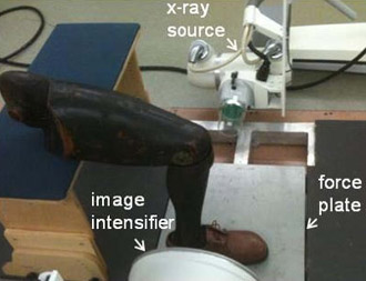
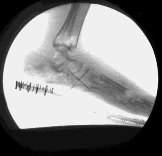
This project is structured to develop a unique biplanar (3-D) fluoroscopic imaging system for in vivo motion analysis of the foot and ankle during walking.This imaging technology will allow real-time assessment of internal bony motion during ambulation.This technology will be coupled with an existing segmental foot and ankle model (Milwaukee Foot Model) for assessment of segmental hindfoot kinetics. Anticipated development results include new knowledge of in vivo bony motion in the foot during ambulation; better information for future footwear; and orthotic and prosthetic development.
- PI
- Taly Gilat Schmidt, Ph.D.
- Patient population
- Other: 5 (children aged 10 – 18 yrs.)
- Project goal
- Develop a biplane x-ray fluoroscopy system for analyzing in vivo motion of the bare or shod foot
D3 Update: May 2013
Presentation by Alex Griffin at GCMAS Annual Conference
INTRODUCTION
Most current gait analysis techniques rely on external markers to quantify segmental foot and ankle motion. Because of the use of external markers, multi-segmental foot models are also unable to accurately analyze the shod foot. This presents a significant challenge to the clinician and engineer interested in quantifying foot kinematics during the shod condition. Those interested in quantifying barefoot motion face similar challenges with the use of external markers, which are subject to motion artifact, model repeatability and external validation requirements, and rigid body motion assumptions. Fluoroscopic analysis, by comparison, eliminates the need for external markers by utilizing images of in vivo bony geometry to create segment/coordinate systems. This process circumvents soft tissue artifact error, and allows for analysis of the shod foot, providing more relevant clinical data to assist in the assessment of orthotics and footwear.
Audio Powerpoint:
D3 Update: 2012 Annual Meeting
- Progress
- Challenges & Solutions
- Accomplishments
- Opportunities
- Plans for Year 3
D3 Update: October 2012
Poster presentation by Alex Griffin at the annual Medical College Research Day.
D3 Specific Aim
- To develop a prototype biplane fluoroscopy system and manual feature extraction method for in vivo studies of subtalar hindfoot kinematics
D3 Conceptual Model
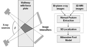
- Proposed 3D biplane fluoroscopy system will be constructed by adding a second gantry to our existing 2D system
- Proposed biplane fluoroscopy system and analysis method
- 3D localization is possible in the region irradiated by both gantries
- Elevated walkway contains a force plate to enable kinetic analysis
- Dynamic biplane x-ray images will be acquired during ambulation
D3 Tasks
- Phantom studies to quantify localization accuracy
- Biplane fluoroscopy hardware and software development
- Dynamic phantom studies to validate biplane localization accuracy
- Pilot studies with 5 children age 10–18 yrs.
D3 Anticipated Timeline
| Activity | Year 1 | Year 2 | Year 3 | Year 4 | Year 5 | |||||||||||||||
|---|---|---|---|---|---|---|---|---|---|---|---|---|---|---|---|---|---|---|---|---|
| Phase 1: Phantom studies with 2D fluoroscopy system | Q1 | Q2 | Q3 | Q4 | ||||||||||||||||
| Phase 2: Construct biplane fluoroscopy system | Q1 | Q2 | Q3 | Q4 | Q1 | Q2 | Q3 | Q4 | ||||||||||||
| Phase 3: Develop feature extraction software | Q1 | Q2 | Q3 | Q4 | Q1 | Q2 | Q3 | Q4 | ||||||||||||
| Phase 3: Dynamic phantom study | Q1 | Q2 | Q3 | Q4 | ||||||||||||||||
| Phase 3: Pilot patient studies (5 children aged 10–18) | Q1 | Q2 | Q3 | Q4 | ||||||||||||||||


